
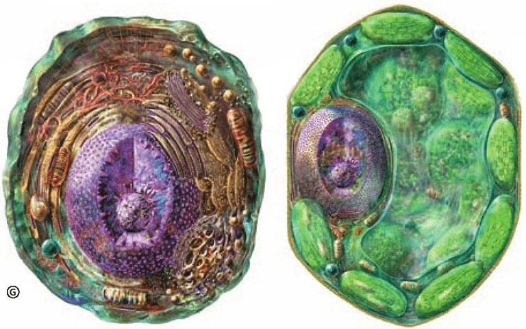
What do cells look like? Before the invention of the microscope, no one had ever heard of a cell, let alone seen one.
More than 300 years ago, Robert Hooke, an Englishman, succeeded in building a microscope that could see tiny objects. When he used the microscope to examine the extremely thin pieces of cork cut with sharp blades, he was fascinated by what he saw: the cork was densely packed with small "empty rooms" like well-made honeycombs. He named these "small rooms""" Cells ", which translated them into" Cells "in Chinese. But all Hooke saw was a cavity surrounded by plant cell walls, and the internal cytoplasm had long disappeared.
Gru, who was also a Briton, further used a microscope to carefully observe the plant slices and drew more than a hundred cell observations. He was also the one who discovered that cells were not empty cavities, filled with juice; cells were not loose individuals that were not interconnected with each other, but closely adhered to each other and had a very precise structure.
Unfortunately, in the 150 years after Robert Hooke discovered cells, humans have never been able to deeply understand cells due to the backward microscope making process.
Until 1839, the Czech and Slovakia histologist Purkinje named the colloidal liquid filling cells protoplasm. However, this name was not recognized by contemporary scholars until the mid-19th century, when the German botanist Moore adopted the term and collectively referred to various types of cell internal substances as protoplasm. From then on, the unity of all the contents within the cell wall is called protoplasm.
Although cells vary in size and shape, they share a common infrastructure. The general structure of all eukaryotic cells is similar: the outermost layer is the cell membrane, and the outer side of the cell membrane of plant cells has an additional layer of cell wall; the interior of the cell membrane is the cytoplasm, and the center of the cell is the nucleus; there are a variety of unique functions distributed in the cytoplasm. Organelles are responsible for biological activities such as nutrient decomposition, synthesis, absorption, and excretion.
The cell nucleus was discovered by British botanist Brown in 1831. It is a dense ball, mostly located in the center of the cell, with a round or oval shape; the nucleus is surrounded by a nuclear membrane, and there are small holes in the membrane that can communicate with the external cytoplasm.
In 1879, German cytologist Fleming used red dye to color small particles in the nucleus. He called this kind of nuclear substance chromatin. In 1882, Fleming again observed that when cells divided, chromatin shrank into thin filaments; in 1888, German anatomist Waldel named chromatin condensed into a rod as chromosomes, that is, the essence of chromosomes is folded and condensed chromatin.