
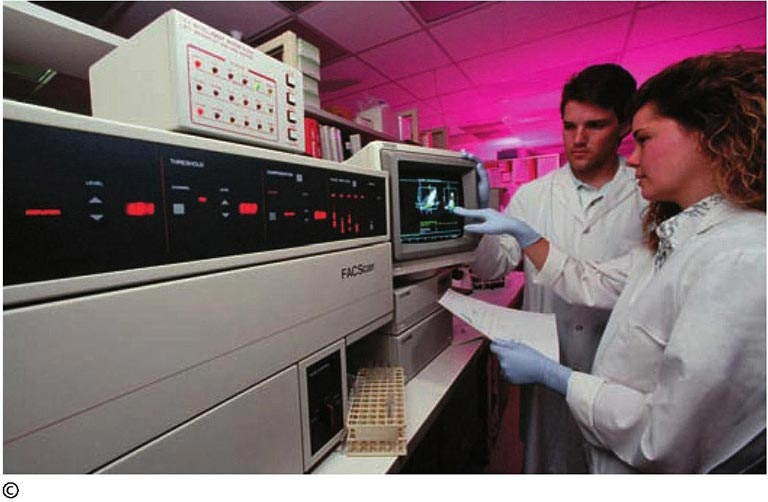
The number of stem cells in the human body is very small, only about one thousandth of the cells of various tissues. Under the microscope, the "appearance" of stem cells is similar to other cells. So how can scientists accurately discover stem cells from the ocean of cells? How to separate them? How to get them to "report in and out of the queue"?
It turns out that the proteins embedded on the surface of each cell are different, and stem cells are no exception. These proteins are like badges worn by people that can be used to identify a person. But these "badges" are too small to be seen with a microscope. So what should we do? Scientists take advantage of the property of antibodies that specifically bind to antigens, use specific antibodies to "label" the "badges" of stem cells, and then attach fluorescent groups to the antibodies. Under the irradiation of laser light, the fluorophore will emit fluorescence. Because tissue cells do not have the unique badge of stem cells, the antibodies connected to the fluorophore cannot bind to it and cannot emit fluorescence.
Next, let this group of majestic mixed cells pass through a thin tube in turn. This special "wooden bridge" is very narrow, allowing only one cell to pass through at a time. As the fluorescent cells pass through the thin tube, the machine charges them. Behind the thin tube is an electric field. Because stem cells are charged, their path will be deflected and enter a special collection tube. Other cells were not labeled, were not charged, and did not deflect in the electric field and entered other collection tubes. In this way stem cells are isolated from all types of cells.