
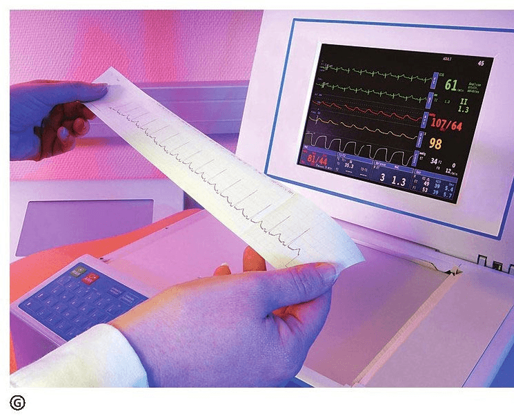
Why does the heart produce an electrocardiogram The generation of electrocardiograms starts with the bioelectrical phenomenon. This phenomenon was discovered very early, but it didn't take long to truly understand it.
Aristotle, an ancient Greek scholar in the 4th century BC, observed that when catching food, the lightning rays first applied electric shocks to animals in the water, paralyzing them. The ancient Greeks and ancient Romans used electric shocks from black lightning rays to treat wind pain and headaches. But it was not until the establishment of electricity in the 18th century that people gradually understood the nature of animal discharges. In 1786, Italian doctor Galvani discovered that if a circuit composed of two metals was used to connect the neuromuscles of a newly prepared frog, the muscles would immediately twitch and shake. He pointed out that this was because the neuromuscular tissue had an intrinsic form of current. However, Italian scientist Volta believed that the shaking of frog's legs was caused by the contact potential difference, and established the theory of electromotive force in metal contact, thus inventing a Volta battery that can generate stable current. The two men argued for a long time about the reason for the frog's legs to shake.
In 1842, Italian physiologist Matteucci proved that the contraction of frog hearts was accompanied by current, which finally settled the debate between Galvani and Volta, and developed cardiac electrophysiology. Scientists have conducted a long-term exploration of the electrical activity of the heart. In 1908, Dutch doctor Eintopine proved the influence of heart rate and respiration on electrocardiogram and proposed that electrocardiogram could be used for clinical diagnosis. After World War II, with the rapid development of electronic instruments, electrocardiograms were used on a large scale in medicine.
More than 200 years of exploration have shown that electricity is ubiquitous in living things. The essence of the life process is the electron transfer process, especially energy conversion, nerve conduction, photosynthesis, and breathing processes. An electrocardiogram is just one of the most common examples.
Let us explain the formation process of electrocardiogram from micro to macro. Cardiomyocytes play a key role in the beating heart. Its cell membrane separates the cytoplasm and extracellular matter, and the embedded proteins on the cell membrane can selectively pass or even actively carry some ions, such as potassium ions, sodium ions, etc., which are charged. This channel protein opens or closes ion channels through changes in the shape of the protein, which means that it can control whether certain ions are allowed to pass through.
Electrocardiogram examination Due to the different permeability of myocardial cell membranes to different ions such as potassium and sodium, the distribution of different ions inside and outside the cell membrane is different. When the cell is resting, the difference in the distribution of these charged ions causes the potential outside the membrane to be higher than that inside the membrane; When local membrane is excited, the permeability of the channel protein to ions will change, and the ions inside and outside the membrane will redistribute, resulting in a lower potential outside the membrane than inside the membrane. This excitement can be transmitted along the cell membrane, causing the cell membrane to excite in a spatial order, and thus the potential also differs and changes in a certain spatial order. When the myocardial cell membrane is excited, biochemical changes occur within the tissue through a mechanism called excitation-contraction coupling, which converts electrical energy into mechanical energy, thereby shortening the muscle fibers.
Excitation of cardiomyocytes throughout the heart is temporal and spatial, resulting in asynchronous contraction of the myocardium. Only in this way can the heart complete its blood pumping function in an orderly manner. The precise conduction of cardiac excitability depends on the fast-conducting fibers in the heart. At a certain moment, a certain part of the heart is in a state of excitement and contraction, and the rest of the heart is in a state of relaxation. Over time, the parts that contract and relax also change. When we place electrodes on specific parts of the body (such as the right arm and left leg), we can record the overall difference in the electrical potential that reflects excitement in different parts of the heart, which is the electrocardiogram we see.
If the potential conduction mechanism within the heart fails, or a certain part of the myocardium is damaged, this overall potential change pattern will change and is reflected in the electrocardiogram. Therefore, electrocardiogram examination can be used to diagnose a variety of heart diseases and save precious lives.